Advancing Breast Imaging: Exploring Digital Breast Tomosynthesis
Introduction
Breast cancer remains one of the most common cancers affecting women worldwide, and early detection is critical for improving survival rates and treatment outcomes. Over the years, advancements in breast imaging technology have played a vital role in enhancing diagnostic accuracy and reducing unnecessary biopsies. Among the most significant breakthroughs in recent decades is Digital Breast Tomosynthesis (DBT) – often referred to as 3D mammography. This cutting-edge imaging technique is transforming how radiologists detect and evaluate breast abnormalities, leading to better patient care and outcomes.
Definition
Digital Breast Tomosynthesis (DBT) is an advanced imaging technique used in breast cancer screening that creates a 3D reconstruction of the breast by taking multiple X-ray images from different angles. This technology improves the detection of abnormalities by reducing tissue overlap seen in traditional mammography, allowing for clearer and more accurate visualization of breast tissues.
Advantages of Digital Breast Tomosynthesis
Improved Cancer Detection Rates:
Detecting breast tumours that 2D mammography might miss is one of DBT’s most important advantages. Studies show that DBT increases cancer detection rates by 20-40%, particularly for invasive cancers. The 3D images help differentiate real abnormalities from normal overlapping tissue, allowing for earlier and more accurate diagnosis.
Reduced False Positives and Recall Rates:
Breast cancer screening false positives might result in needless worry, further imaging, and biopsies. DBT lowers the quantity of recalls and false alerts by delivering sharper images. Research indicates that DBT can reduce recall rates by up to 40%, meaning fewer women have to return for additional testing without a definitive reason.
Better Visualization in Dense Breasts:
Women with dense breast tissue pose a diagnostic challenge because dense tissue can mask tumors on 2D mammograms. Through its ability to penetrate dense tissue, DBT’s 3D imaging enhances cancer detection in these patients and yields more accurate screening findings.
Enhanced Diagnostic Confidence:
Radiologists reviewing DBT images report greater confidence in their interpretations. The ability to view breast tissue in thin slices reduces uncertainty and supports more precise clinical decisions, which translates into better patient management.
Who Should Consider Digital Breast Tomosynthesis?
While DBT is becoming the standard of care in many breast imaging centers, not all facilities offer it yet, and insurance coverage can vary.
Screening for Average-Risk Women:
Many experts recommend DBT as the preferred screening method for women over 40 or those starting routine mammography screening. Its improved detection and reduced recall rates make it a compelling option for early breast cancer detection.
Women with Dense Breasts:
Since dense breast tissue lowers the sensitivity of traditional mammography, DBT is highly beneficial for these women. In fact, some states in the U.S. require radiologists to inform women about their breast density and the availability of DBT.
Women at Higher Risk:
Women with a family history of breast cancer or other risk factors may benefit from the enhanced imaging detail provided by DBT, although additional modalities such as MRI or ultrasound may also be recommended.
The Procedure: What to Expect During a DBT Exam?
DBT exams are very similar to traditional mammograms in terms of procedure and experience. Here’s what typically happens:
- The patient stands in front of the DBT machine.
- The technologist positions the breast on the imaging platform and compresses it gently but firmly to obtain clear images.
- Multiple photos from various angles are captured in a matter of seconds by the X-ray arm moving in an arc around the breast.
- The compression may be a bit uncomfortable but usually only lasts for a short time.
- The entire exam typically takes about 10 to 15 minutes.
Because DBT uses low-dose X-rays, the radiation exposure is slightly higher than a standard mammogram but still within safe limits recommended by regulatory agencies.
Challenges and Considerations with Digital Breast Tomosynthesis
While DBT offers numerous advantages, there are some considerations to keep in mind:
- Radiation Dose: The radiation dose in DBT is slightly higher than standard 2D mammography but remains low and safe. Some systems combine DBT with 2D images, which can increase dose, though synthetic 2D images can reduce the need for additional exposures.
- Cost and Accessibility: DBT equipment is more expensive than traditional mammography machines, and not all centers have upgraded. This can affect patient access, particularly in rural or underserved areas.
- Insurance Coverage: While many insurers cover DBT, coverage policies vary, and out-of-pocket costs may apply.
- Learning Curve: Radiologists require training to interpret DBT images accurately, which can affect availability and turnaround time.
The Future of Breast Imaging: Integration and Innovation
Although digital breast tomosynthesis marks a significant advancement in breast imaging, innovation will continue. Researchers and clinicians continue to develop new technologies and methods to further improve breast cancer detection, including:
- Artificial Intelligence (AI) and Machine Learning: AI algorithms are being developed to assist radiologists in interpreting DBT images, improving accuracy, and reducing workload.
- Contrast-Enhanced Tomosynthesis: Combining DBT with contrast agents to highlight blood flow and tumor angiogenesis for better characterization of lesions.
- Automated Breast Ultrasound (ABUS): Used in conjunction with DBT for enhanced imaging in women with dense breasts.
These advancements promise to make breast cancer screening more precise, personalized, and accessible in the coming years.
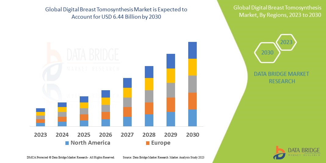
Growth Rate of Digital Breast Tomosynthesis Market
According to Data Bridge Market Research, the digital breast tomosynthesis market, valued at USD 2.34 billion in 2022, is projected to grow at a compound annual growth rate (CAGR) of 13.5% from 2023 to 2030, reaching USD 6.44 billion.
Learn More: https://www.databridgemarketresearch.com/reports/global-digital-breast-tomosynthesis-market
Conclusion
Digital breast tomosynthesis is transforming breast imaging by producing 3D images of breast tissue that are more detailed and clearer. Its ability to improve cancer detection rates, reduce false positives, and enhance visualization in dense breasts makes it an invaluable tool in the fight against breast cancer.